The spinal column comprises three main components: the vertebrae, the intervertebral discs, and the muscles/tendons/ligaments that support them. Between the two vertebrae, there is a cushion-like organ called intervertebral disc that made up of a fibrous wall and a gelatinous nucleus. The connection of the vertebrae and discs creates a channel inside the spine through which the spinal cord and nerve roots pass. The nerve roots then exit the spine through a hole called the neuro-foramen and progress into the peripheral organs.
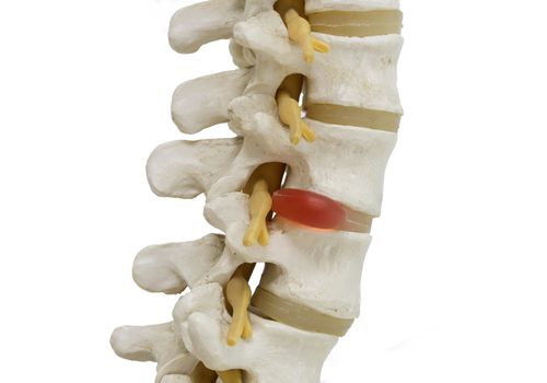
The spinal column is categorized from top to bottom into five groups based on the areas where they locate. These areas include seven cervical vertebrae, twelve dorsal vertebrae, five lumbar vertebrae, four fused sacral vertebrae, and four to five fused caudal vertebrae. However; in a small percentage of ordinary people, some vertebrae move between these areas.
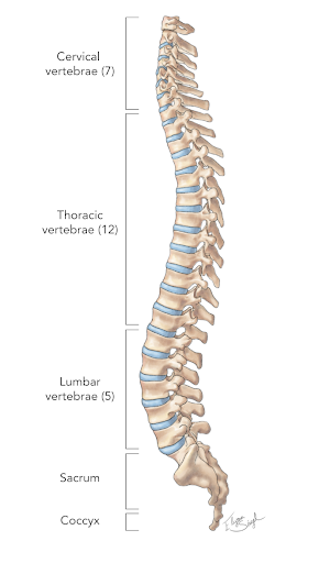
The cervical and lumbar vertebrae serve crucial roles as the nerve roots coming out of these areas form complex neural plexus that are responsible for delivering nerves to the upper and lower limbs.
Compression of the discs in these areas can lead to protruding of the nucleus. This phenomenon called discal hernia can cause mechanical and chemical damage to adjacent nerve roots that cause pain, numbness, and other neurological symptoms in the limbs.
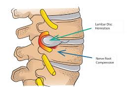
In these cases, with relative rest, anti-inflammatory and analgesic drugs, the injury can be controlled. If the injury is not critical, the condition can gradually improve. In cases of severe progressive damage to the spinal roots, surgery or similar interventions can be helpful.
You may find proper exercise for this illness in the Rehabex app.
Prepared by Rehabex team.

